See at two scales
Three contextual cameras guide motion across the workspace. A fixed-field stereoscope view supports focusing, centering and final alignment.
ICRA 2026 · CO-FIRST AUTHOR
Automating microscale device placement with visual feedback and accessible laboratory hardware.
01 · THE SYSTEM
Placing a tiny implantable device requires coordinated motion at a scale that is difficult to handle manually. This work brings cameras, micromanipulators and an injector together into an automated laboratory system built with commercial off-the-shelf components.
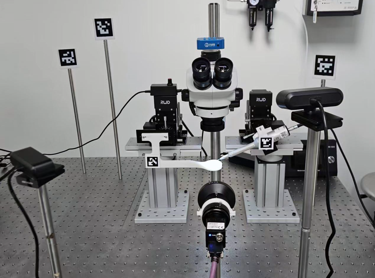
Three contextual cameras guide motion across the workspace. A fixed-field stereoscope view supports focusing, centering and final alignment.
The two micromanipulators move the sample and injector. Their control interface does not provide direct position readback, so the system uses visual feedback to guide motion.
A custom biocompatible, 3D-printed holder supports the sample and provides visible features for locating its center.
02 · THE WORKFLOW
After the operator places the prepared sample holder on the stage, four stages progressively align the sample and the device-loaded pipette.
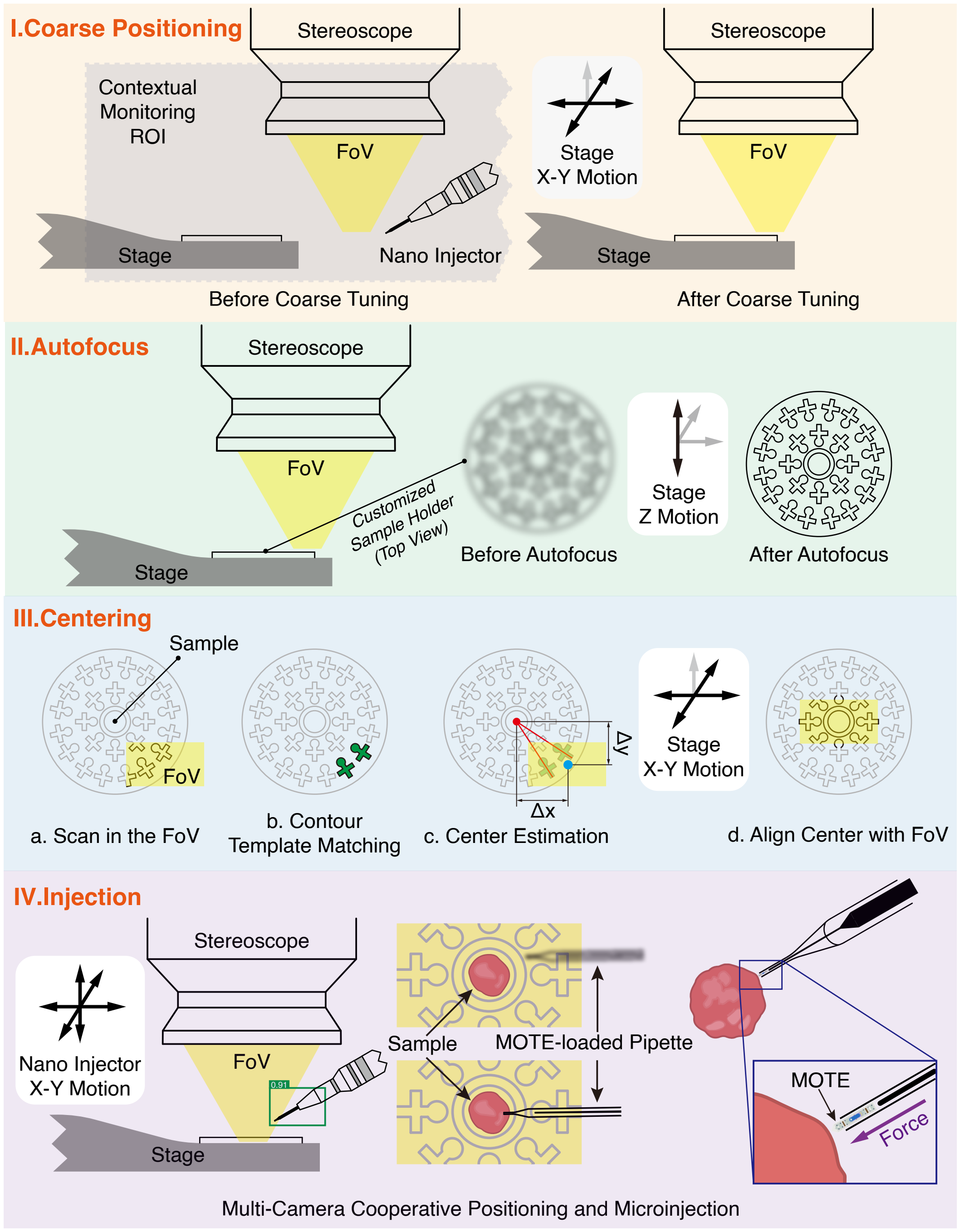
Contextual camera views and ArUco markers estimate the stage pose and move the holder into the stereoscope’s field of view.
The system adjusts the sample position using image feedback to bring it into focus without motorized microscope zoom or focus.
Features on the sample holder are matched to estimate its center, then aligned with the camera view.
Object detection and segmentation locate the pipette tip and gel. Visual contact detection stops the approach, then the nano injector advances the device with a silver wire.
03 · UNDER THE MICROSCOPE
A microscale optoelectronic tetherless electrode (MOTE) is loaded into a pulled glass pipette and delivered into a tissue-mimicking agarose sample. The sequence below shows the injection and subsequent withdrawal steps.
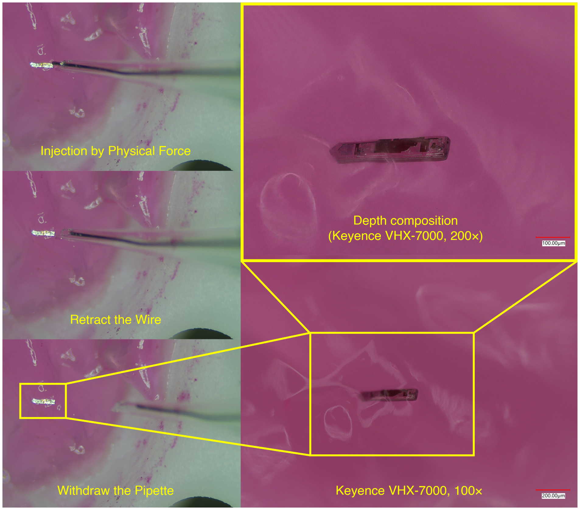
Under optical illumination, the implanted MOTE emitted the expected pulses during the post-injection check. This confirms that the device remained functional in the gel; the experiment does not record biological neural signals.
04 · EVALUATION
The study reports 30 automated injection trials using two MOTEs, alternated and reused after each injection.
The average times below sum to 158.2 seconds, or about 2.6 minutes. The paper compares this with an experienced user’s manual procedure of approximately five minutes.
| Stage | Time (seconds) |
|---|---|
| Stage alignment | 30.5 |
| Autofocus | 24.2 |
| Centering | 56.3 |
| Injection | 47.2 |
| Sum of reported stage averages | 158.2 |
The system was also demonstrated with a 008004 surface-mount capacitor measuring 250 × 125 × 125 µm. The experiments use tissue-mimicking agarose, and loading devices into the pipette still requires preparation. The reported time reduction is an efficiency result, not an injection success rate.
PUBLICATION
ICRA 2026
† Equal contribution. The official ICRA 2026 title is shown above; figures and detailed results follow the research manuscript.